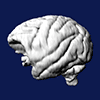
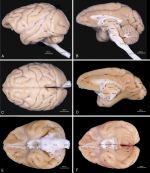
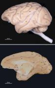
The atlas consists of views of the external surfaces of
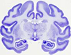
mulatta brains and images of the Nissl-stained histology
of three serially sectioned brains.
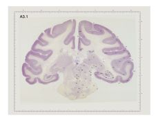
All of the brains were from adult monkeys, perfused with
paraformaldehyde. The brains to be sectioned had electrodes
introduced in the frontal, parasagittal or horizontal
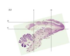
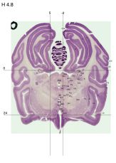
planes and at known Horsley-Clarke coordinates in order to
provide fiduciary marks for alignment, registration and
coordinate identification. A grid showing coordinates in mms
has been overlain on each section. The coronal atlas is now
finalized; the sagittal and horizontal atlases are works
in progress. Labeling of structures is the same as in the
high resolution images found under the Macaca mulatta
datasets. Section numbers are the same as in the high
resolution datasets so that sections can be matched for
higher resolution viewing.